Automatic Detection of Pathologies in Conventional Scanner Data
Détection automatique d’anomalies sur examens tomodensitométriques standards
Philips Research France · CREATIS (INSA Lyon, Université Claude Bernard Lyon 1) · Hospices Civils de Lyon
Abstract
Computed tomography (CT) is a common medical imaging examination used to diagnose or monitor many conditions, particularly in the thoracic region. These CT scans produce large amounts of high-quality volumetric data. However, the increasing number of scans and images produced by acquisitions result in a significant workload for radiologists. Radiologists primarily focus on suspected anomalies based on the initial clinical indication, which may lead to underdiagnosis of other atypical or unrelated anomalies. This underscores the importance of developing automatic alert systems based on AI to assist in identifying and locating incidental pathologies. Our thesis work focused on developing this type of system, applied to the detection and characterization of pulmonary pathologies in CT scans, using machine learning methods that require minimal annotations and are independent of clinical indications.
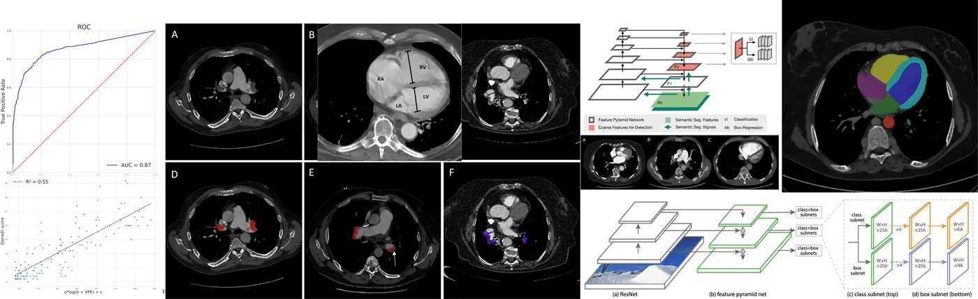
In this thesis, we explore different supervision methods to gradually reduce the dependency on the number of annotated data needed to train anomaly detection algorithms in CT scans. Through supervised learning, we proposed a method for detecting pulmonary embolisms in scans based on the segmentation of blood clots responsible for the embolism. We compared this segmentation-based supervised approach to a classification approach supervised only by the indication of the presence of a pulmonary embolism, illustrating the impact of the type of supervision on performance. We also proposed methods for automatically estimating clinical biomarkers such as the Qanadli score and the VD/VG ratio.
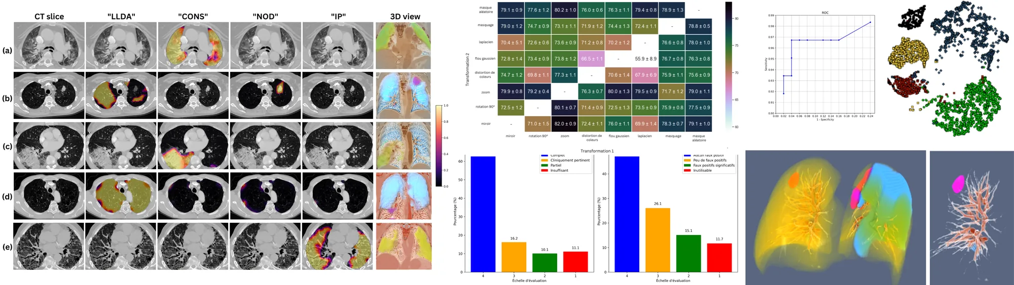
Although this supervised approach works very well and is often preferred for developing clinical decision support systems specific to a pathology, an anomaly detection system must support all pathologies and anatomical conditions encountered, and we do not currently have a representative annotated database. Therefore, we opted for a semiological approach that identifies specific patterns and morphologies present in most pulmonary pathologies (emphysema, cysts, nodules, consolidation, and interstitial patterns) and proposed a weakly supervised approach based on contrastive self-supervised pre-training. This approach allows detecting and locating a wide range of anomalies without requiring exhaustive and detailed data annotation.
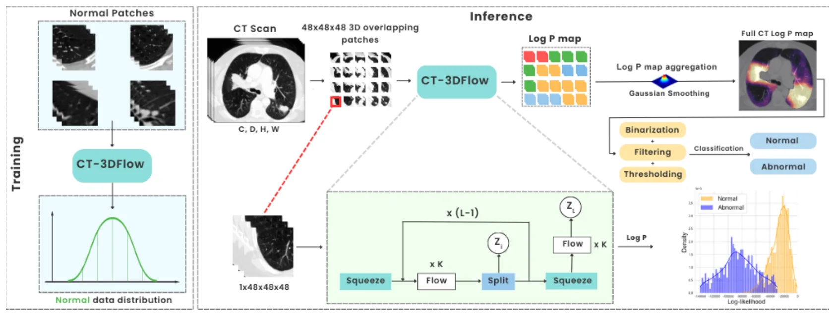
Finally, we proposed an unsupervised approach using a generative method. Generative methods allow learning and representing complex distributions ; we learned the distribution of "normal" patients to then identify anomalies by deviation from this distribution. Training this model only requires a set of examinations from patients without anomalies, thus overcoming the difficulties in creating a representative database of all observed pathologies.
Keywords: Anomaly detection · Computed Tomography · Supervised learning · Weakly supervised learning · Unsupervised learning
Objectives
The thesis focuses on chest CT: a widely used modality with many examinations and highly reproducible volumetric data. The goal is a reading assistant that detects and localizes thoracic anomalies on any 3D CT scan with little annotation, by progressively reducing the level of supervision, from supervised to unsupervised methods.
Table 1: Three levels of supervision explored in the thesis, from most to least annotation.
| Part | Supervision | Target anomalies | Annotation |
|---|---|---|---|
| I | Supervised | Pulmonary embolism | Pixel level (lesion segmentation) |
| II | Self- and weakly supervised | 5 common semiological signs: emphysema and cysts, nodules, consolidation, interstitial pattern, normal | Patch level (image regions) |
| III | Unsupervised | Any deviation from normal lung anatomy | None (normal scans only) |
I. Supervised: pulmonary embolism
Pulmonary embolism (PE) is a frequent and serious condition in which a clot blocks the pulmonary arteries. Diagnosis relies on CT angiography, the RV/LV diameter ratio and the Qanadli obstruction score. We compared two levels of supervision: a 3D segmentation network trained on fine clot segmentations from 54 PE-positive patients, and a 2D CNN + RNN classifier trained on image-level labels from more than 7,000 patients.
- Segmentation beats labels: learning from a few precise clot segmentations outperformed classification trained on thousands of image-level labels.
- Severity, not just presence: the same pipeline estimates the Qanadli score and the RV/LV ratio automatically.
- First place at the 2022 JFR data challenge (Journées Francophones de Radiologie) organized by the French Society of Radiology.
Full write-up: Detection and severity quantification of pulmonary embolism.
Supervised learning works very well for a specific pathology, but an anomaly detection system must cover every pathology and anatomical condition, and no representative annotated database exists. The next two parts reduce the need for annotation.
II. Self- and weakly supervised: semiological signs
Instead of one detector per disease, a semiological approach targets the patterns shared by most lung diseases: emphysema and cysts, nodules, consolidation and interstitial patterns. A 3D patch classifier is pre-trained with SimCLR contrastive learning on unlabeled patches from 1,337 patients, then fine-tuned on 4,000 visually selected patches from 1,330 patients. At inference, overlapping patches produce per-sign probability maps that are aggregated into a patient-level prediction.
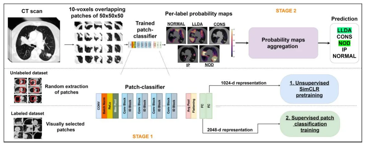
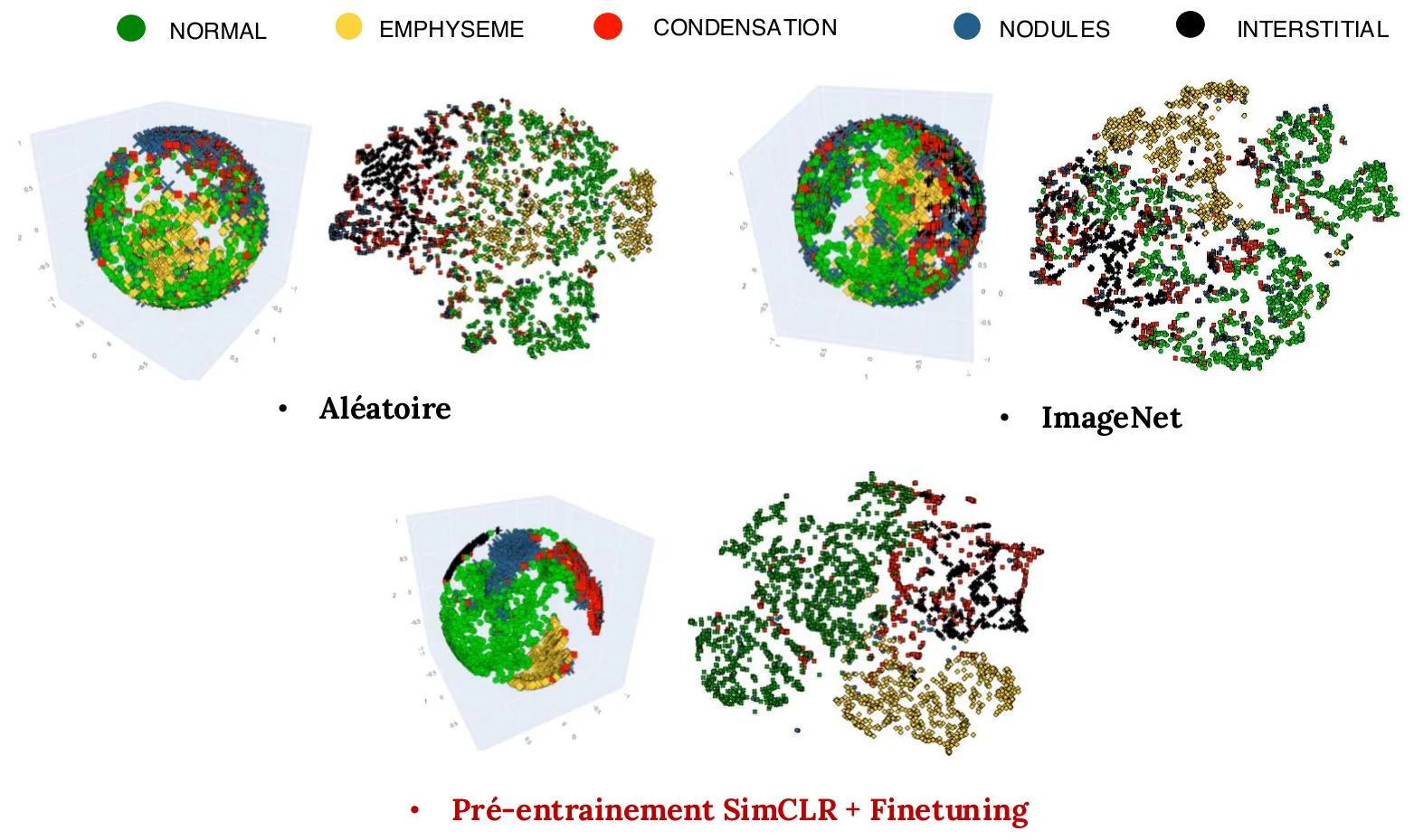
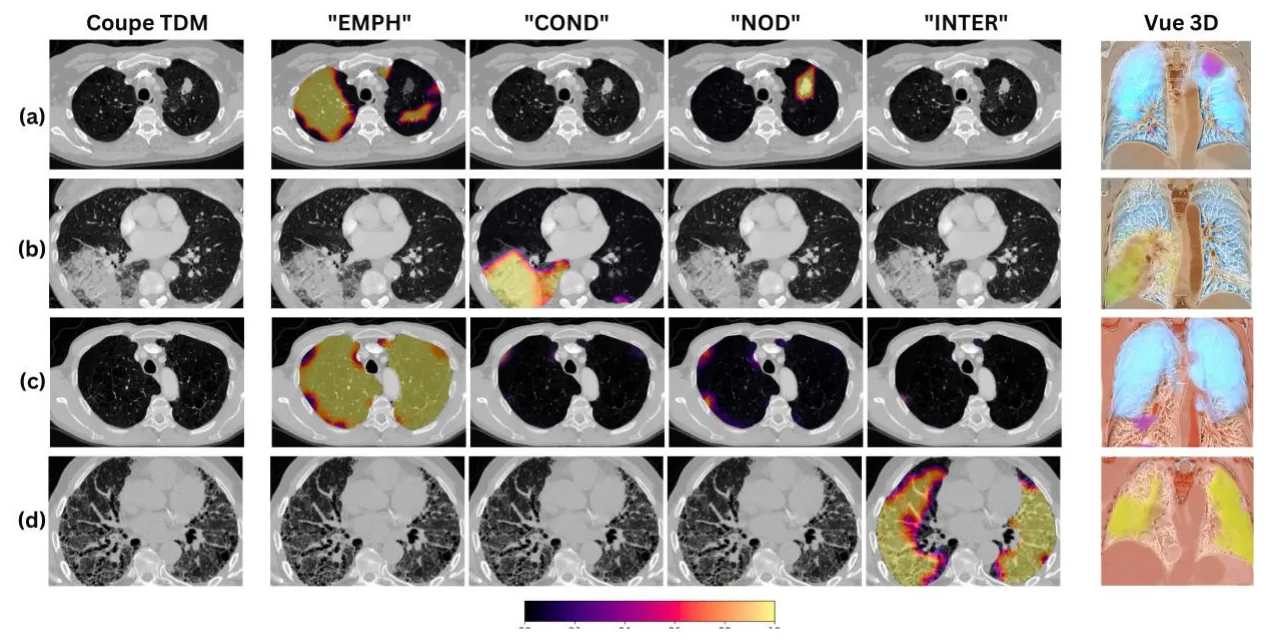
- Self-supervision helps: SimCLR pre-training gave a clear boost in patient-level classification over random and ImageNet initialization, on a 251-patient test set.
- Simultaneous localization: the maps localize several abnormal regions at once, consistent with the ground-truth annotations.
- 88.8% recall and 88.3% precision of localization in a qualitative analysis by an expert radiologist.
Full write-ups: Weakly-supervised pathology detection and localization and Tailored 3D CT contrastive pretraining.
III. Unsupervised: normalizing flows
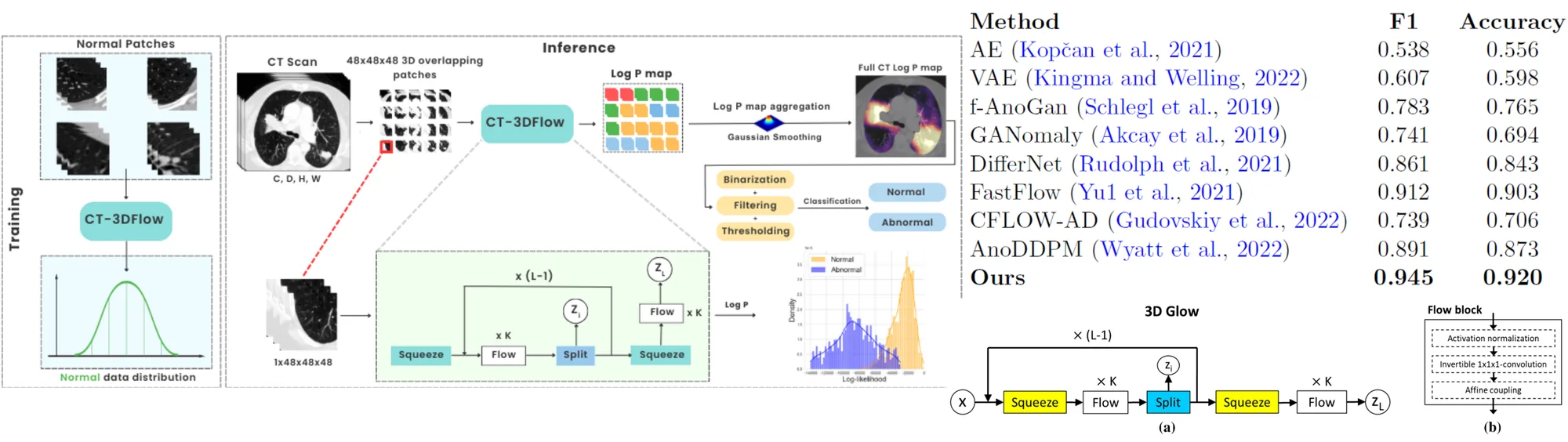
Normalizing flows are generative networks that learn the exact probability distribution of the data by transforming it into a simpler one. Trained on masked lung images from normal patients only, the model assigns low likelihood to anything that departs from normal anatomy. Thresholds and filters are selected on a validation set, and slice- or patch-level scores are aggregated into a patient-level decision.
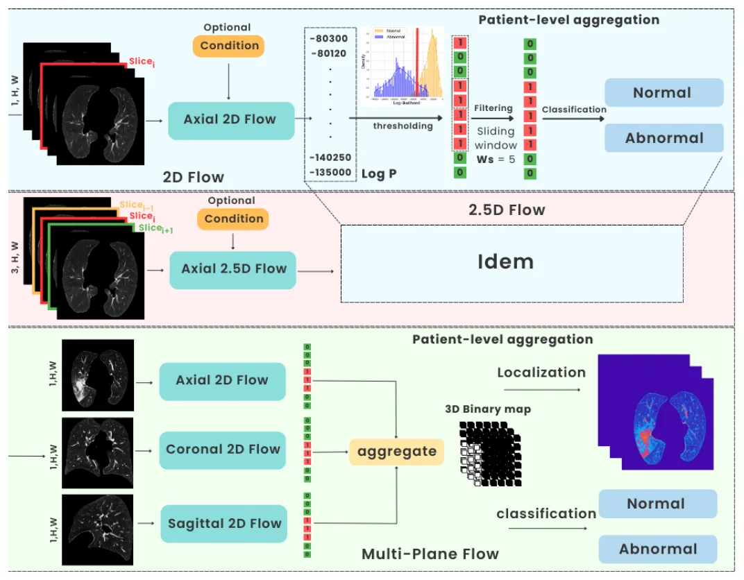
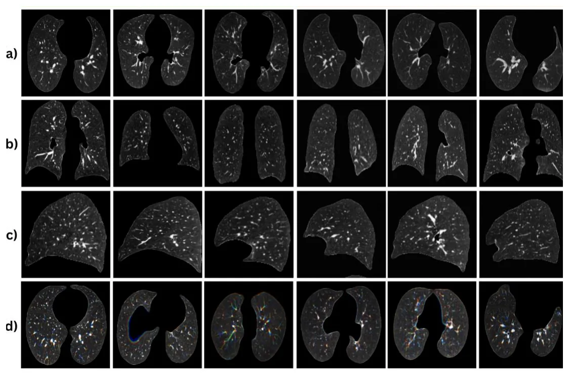
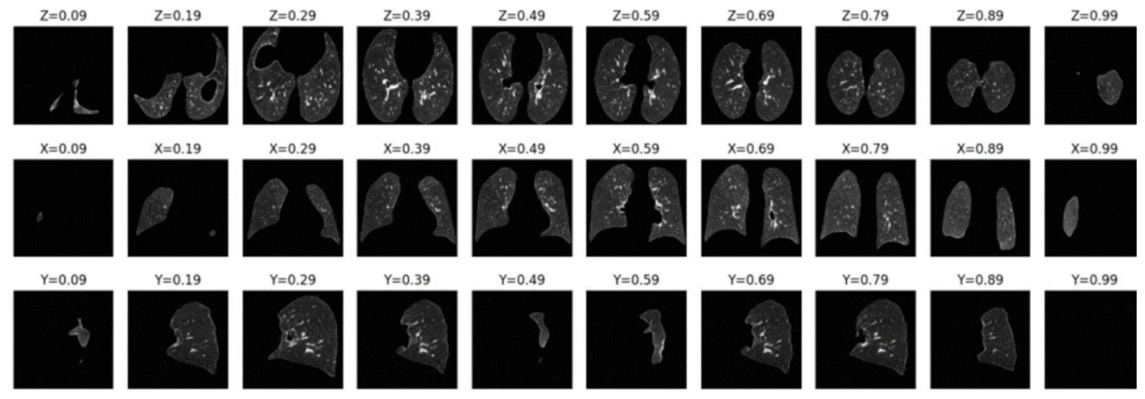
The 3D variant, CT-3DFlow, works on 3D patches to catch pathologies that are hard to see in 2D or 2.5D and to localize them in 3D.
Table 2: Patient-level performance on a test set of 251 patients with various combinations of anomalies (defense slides). Best in bold; best unsupervised F1 and accuracy underlined.
| Method | Family | AUROC | F1 | Accuracy |
|---|---|---|---|---|
| AE | Reconstruction | 0.608 | 0.538 | 0.556 |
| VAE | Reconstruction | 0.647 | 0.607 | 0.598 |
| One-Class SVM | One-class | 0.872 | 0.853 | 0.867 |
| DRAEM | Reconstruction | 0.651 | 0.621 | 0.628 |
| f-AnoGAN | GAN | 0.821 | 0.783 | 0.765 |
| GANomaly | GAN | 0.772 | 0.74 | 0.694 |
| FastFlow | Normalizing flow | 0.942 | 0.912 | 0.903 |
| CFLOW-AD | Normalizing flow | 0.732 | 0.739 | 0.706 |
| DifferNet | Normalizing flow | 0.901 | 0.861 | 0.843 |
| AnoDDPM | Diffusion | 0.924 | 0.891 | 0.873 |
| SimCLR + fine-tuning (Part II) | Self-supervised | 0.963 | 0.974 | 0.952 |
| NF 2D | Unsupervised (ours) | 0.933 | 0.909 | 0.891 |
| NF 2D, conditioned | Unsupervised (ours) | 0.971 | 0.931 | 0.909 |
| NF 2.5D | Unsupervised (ours) | 0.956 | 0.903 | 0.916 |
| NF multi-plane | Unsupervised (ours) | 0.918 | 0.9 | 0.895 |
| NF 3D (CT-3DFlow) | Unsupervised (ours) | 0.952 | 0.939 | 0.924 |
| NF 3D, conditioned | Unsupervised (ours) | 0.927 | 0.902 | 0.9 |
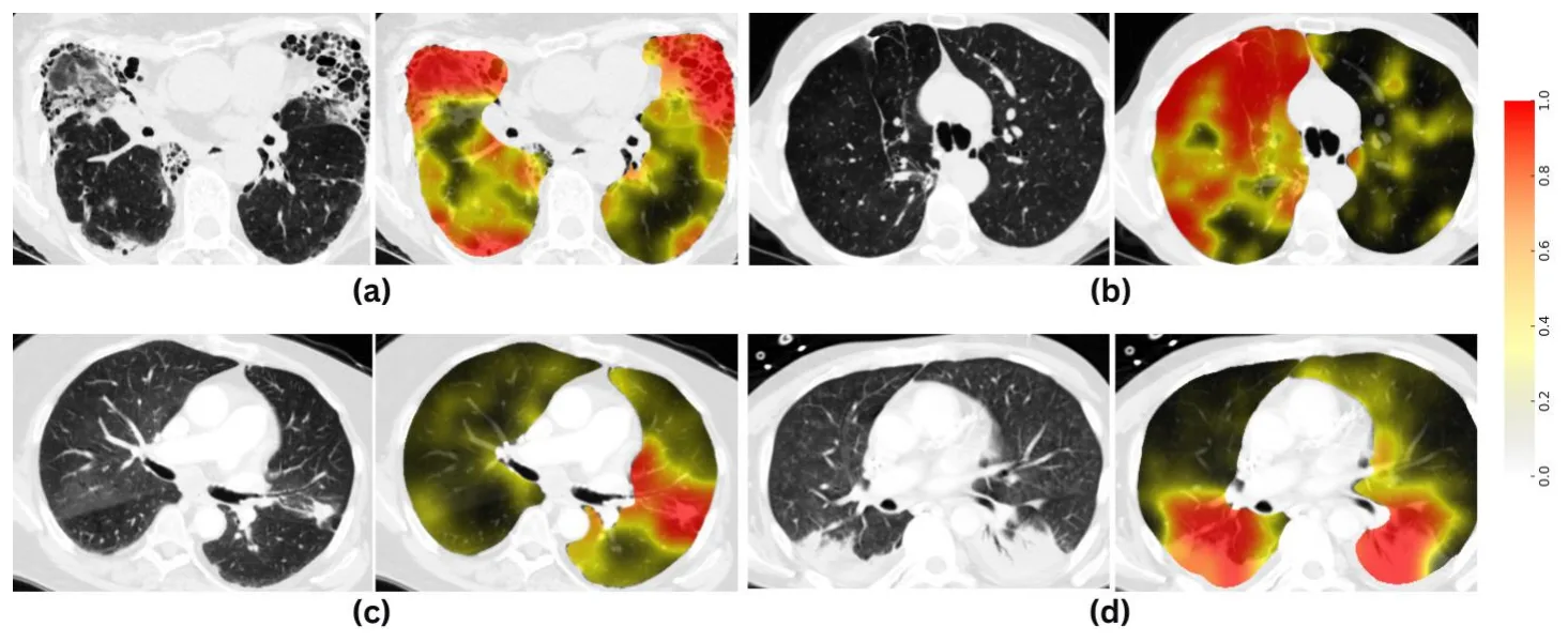
- Beats other unsupervised families: our flows outperform one-class SVM, autoencoders, GANs, diffusion models and other normalizing flows.
- 3D over 2D: 3D models perform better than the 2D versions.
- Close to self-supervision: without any annotation, the flows are competitive with the SimCLR model of Part II.
- Localization without supervision: the likelihood maps highlight the abnormal regions.
Full write-up: CT-3DFlow.
Conclusion & perspectives
- Less annotation: exploring several levels of supervision reduced the dependence on annotations while keeping good performance.
- Self-supervision and semiology: a promising route to generic reading-assistance systems.
- Unsupervised methods: an interesting alternative for anomaly detection, outperforming existing state-of-the-art methods.
- Current limits: performance does not yet allow routine clinical use, but opens promising perspectives.
Next steps include multimodal methods that combine imaging modalities (MRI, ultrasound, PET), text reports and biomarkers, and spectral CT, which adds information on tissue composition by separating materials by their absorption at different energies.
Jury
- Pr. Isabelle Bloch, Sorbonne Université · Reviewer
- Pr. Alexis Jacquier, Aix-Marseille Université · Reviewer
- Pr. Adrian Basarab, Université Claude Bernard Lyon 1 · Examiner
- Dr. Constance de Margerie-Mellon, Université Paris Cité · Examiner
- Dr. Olivier Nempont, Philips Research France · Co-supervisor
- Dr. Alexandre Popoff, Philips Research France · Co-supervisor
- Pr. Loïc Boussel, UCBL, Hospices Civils de Lyon · Thesis director
Publications from the thesis
- Detection and severity quantification of pulmonary embolism with 3D CT data using an automated deep learning-based artificial solution, Diagnostic and Interventional Imaging, 2024.
- Weakly-supervised learning-based pathology detection and localization in 3D chest CT scans, Medical Physics, 2024.
- BERT-based natural language processing analysis of French CT reports: application to the measurement of the positivity rate for pulmonary embolism, Research in Diagnostic and Interventional Imaging, 2023.
- eCAPRI: automated TAVI risk assessment with imaging biomarkers, European Radiology, 2025.
- CT-3DFlow: Leveraging 3D Normalizing Flows for Unsupervised Detection of Pathological Pulmonary CT scans, arXiv, 2024.
- Tailored 3D CT contrastive pretraining to improve pulmonary pathology classification, IEEE ICSP, 2022.
BibTeX
@phdthesis{djahnine2024thesis,
  title  = {D{\'e}tection automatique d'anomalies sur examens
            tomodensitom{\'e}triques standards},
  author = {Djahnine, Aissam},
  school = {Universit{\'e} Claude Bernard Lyon 1},
  year   = {2024},
  number = {2024LYO10167},
  url    = {https://theses.hal.science/tel-05263492}
}