Contrastive Pretraining for Pulmonary Pathology Classification
Learning Representations from 3D CT Patches
INSA Lyon (CREATIS) · Hospices Civils de Lyon · Philips
Abstract
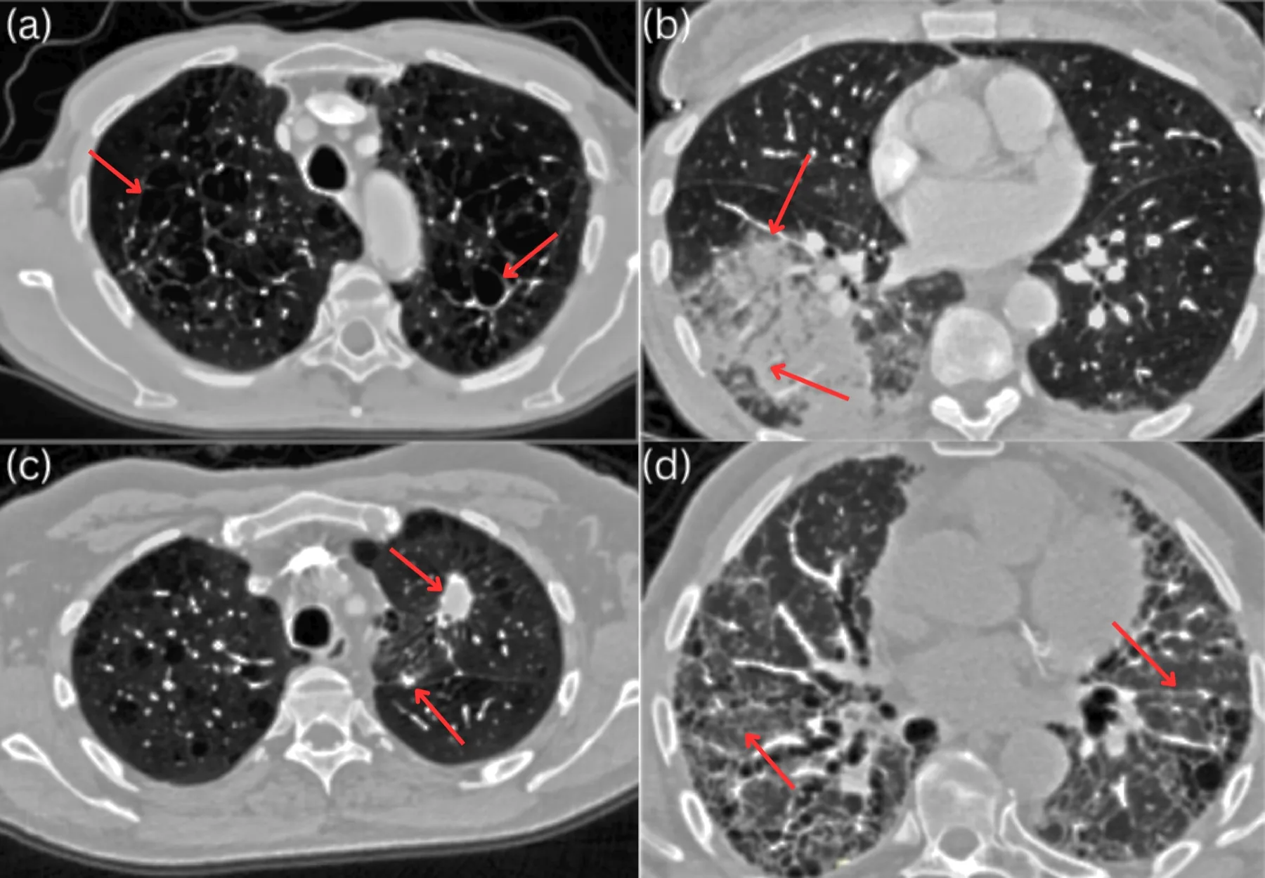
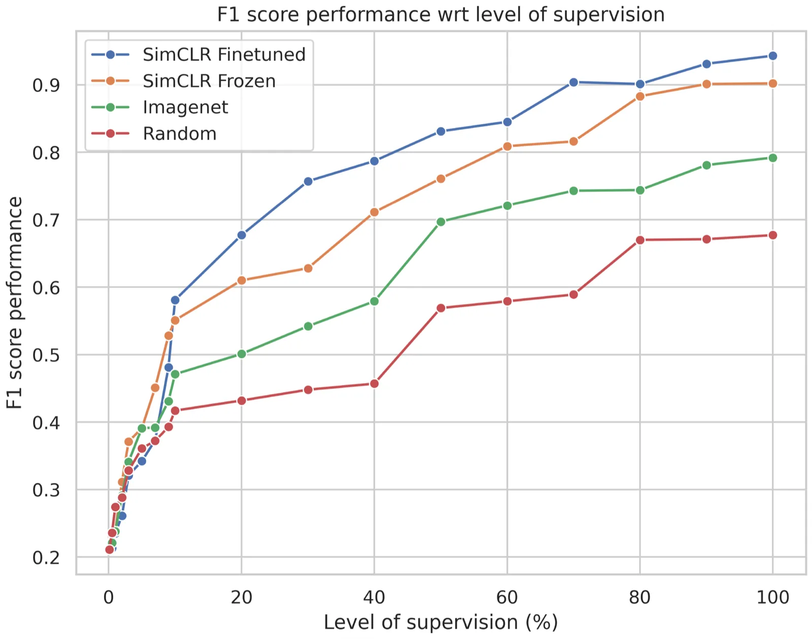
Learning useful representations is a key task for supervised, unsupervised, and self-supervised algorithms. These latter methods have shown great promise for learning meaningful visual representations from unlabeled data, which can then be readily used for downstream tasks (e.g., classification). Recently proposed contrastive self-supervised learning methods have shown high performance on natural images. In this work, we show the usefulness of such approaches in the medical domain when used for 3D chest CT patch pathology classification. We observe that pretraining on unlabeled domain-specific medical images using contrastive self-supervised learning with specific data transformation significantly improves the accuracy of our 3D patch-based pathology classifiers. Specifically, we show that contrastive pretraining outperforms end-to-end supervised training by a large margin (weighted accuracy: 90.33 % vs 77.75 %, respectively).
Keywords: Training; Pathology; Visualization; Three-dimensional displays; Computed tomography; Signal processing algorithms; Self-supervised learning
Method
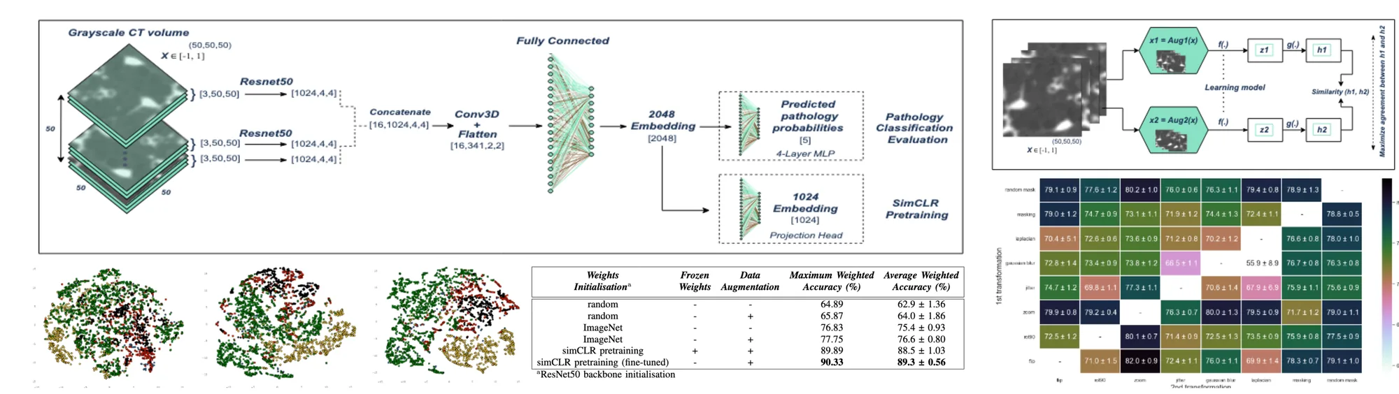
The research overview pairs a CT patch encoder with a SimCLR projection head for pretraining and a classification head for downstream evaluation. It compares transformations and weight-initialization strategies.
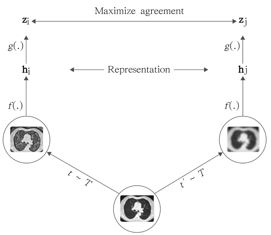
Two transformed views of a CT patch are used during contrastive pretraining. The projection head serves representation learning, while a separate classification head evaluates the learned features. The overview also visualizes the resulting patch embeddings.
2.1SimCLR pretraining
- Patch sampling. Random 50 × 50 × 50 patches containing at least 80% lung.
- Two correlated views. A transformation module randomly alters each patch twice.
- Encoder. CT-Net, a 2.5D alternative to 3D ResNets built on a 2D ResNet-50, producing a 2048-d representation and allowing ImageNet initialization for comparison.
- Contrastive loss. A 2-layer projection head maps representations to 1024-d; the NT-Xent loss pulls the two views together and pushes other patches apart. The head is discarded after pretraining.
2.2Tailoring the transformations to CT
Eight transformations common in medical imaging were evaluated in pairs (56 configurations), each with a frozen pretrained encoder over 10 runs: mirror, 90° rotation, zoom, colour distortion, Gaussian blur, Laplacian, lung masking and a random lung mask from another patient.
Experiments
The research figure compares weight initialization, augmentation, and whether pretrained encoder weights remain frozen. Maximum and average weighted accuracy are reported separately.
Results
Table 1: Classification comparison reproduced from the research overview
| Initialization | Frozen encoder | Augmentation | Maximum weighted accuracy (%) | Average weighted accuracy (%) |
|---|---|---|---|---|
| Random | No | No | 64.89 | 62.9 ± 1.36 |
| Random | No | Yes | 65.87 | 64.0 ± 1.86 |
| ImageNet | No | No | 76.83 | 73.45 ± 0.93 |
| ImageNet | No | Yes | 77.75 | 76.6 ± 0.80 |
| SimCLR | Yes | Yes | 89.89 | 88.5 ± 1.03 |
| SimCLR, fine-tuned | No | Yes | 90.33 | 89.3 ± 0.56 |
The ± values follow the notation in the supplied research figure.
Key findings
- Pretraining: both SimCLR configurations exceed the ImageNet and random-initialization configurations shown.
- Fine-tuning: updating the pretrained encoder yields the highest reported average weighted accuracy.
- Augmentation: the figure also compares CT transformations, showing that the pretraining setup includes choices beyond encoder architecture.
Transformation study
- Zoom matters most: the best transformation pairs all include zoom (mirror then zoom: 82.0% weighted accuracy with a frozen encoder).
- Avoid intensity changes: Gaussian blur, Laplacian and colour distortion degrade accuracy, since Hounsfield values carry the diagnostic signal. The final recipe is random mask, mirror, 90° rotation and zoom.
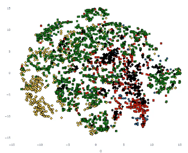
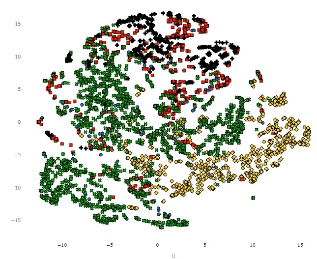
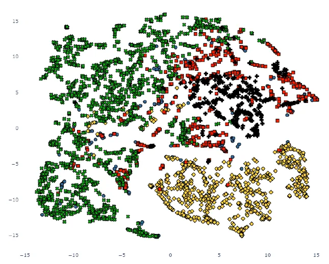
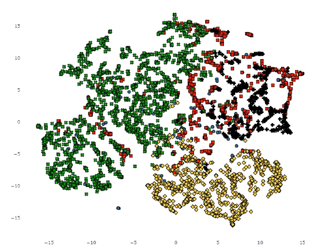
Figure 3: t-SNE projections of test-patch representations. Green NORMAL, blue NOD, orange EMPH, red COND, black INTER. Signs form distinct clusters only with SimCLR pretraining.
Conclusions & limitations
Domain-specific representation learning is the central contribution. The comparison concerns 3D patch classification; it should not be read as a patient-level diagnostic accuracy result.
Related links
Conference paper · Original research overview and comparison table.
BibTeX
@inproceedings{djahnine2022contrastivepretraining,
  title = {Tailored 3D CT contrastive pretraining to improve pulmonary pathology classification},
  author = {A. Djahnine and Popoff Alexandre and E. Jupin-Delevaux and Cottin Vincent and Nempont Olivier and Boussel Loic},
  booktitle = {16th IEEE International Conference on Signal Processing (ICSP)},
  year = {2022},
  doi = {10.1109/ICSP56322.2022.9965218}
}